Image of the Week Gallery
Acidocalcisomes in Trypanosomatids
Media Details
Created 10/5/2004 5:00:00 AM
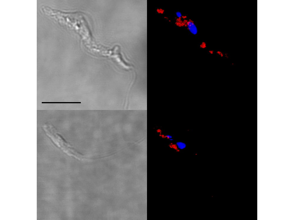
High resolution imaging of acidocalcisomes in trypanosomatids using serial confocal and image reconstruction with VolumeJ. Top panel: Trypanosoma brucei procyclic forms. Bottom panel: Leishmania mexicana amazonensis promastigote forms. Cells were labeled with DAPI (blue) and an antibody specific for acidocalcisomes (red). Both nuclear and extranuclear mitochondrial DNA are well resolved with DAPI. Data was collected on the Leica SP-2 spectral confocal housed at the ITG Microscopy Suite.
Credits
- Peter Rohloff, Ph.D , College of Veterinary Medicine
Visualization Laboratory
Beckman Institute room 2203
405 North Mathews Avenue, Urbana, IL
(217) 300-0566