Image of the Week Gallery
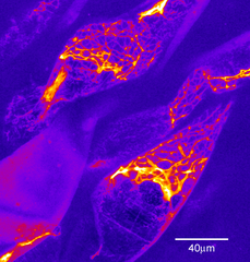
GFP Labeled Microtubules
Media Details
Created 8/28/2003 5:00:00 AM
This image is a composite of 36 sequential optical sections through cells located in the stem of an Arabidopsis thaliana seedling. The structures are microtubules in the plant cells, the cells are expressing a green fluorescent protein conjugated gene product which associates with the microtubules in vivo. The image uses a psueduocoloring technique to allow better discrimination of relative concentrations of fluorescent protein. Noteworthy is the clean 3-D image despite being gathered through a depth of about 15 microns of living plant tissue.
Credits
- Karl Garsha , ITG, Beckman Institute
Visualization Laboratory
Beckman Institute room 2203
405 North Mathews Avenue, Urbana, IL
(217) 300-0566