Image of the Week Gallery
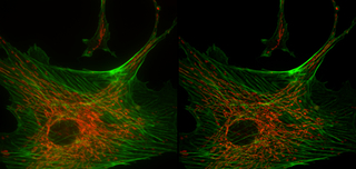
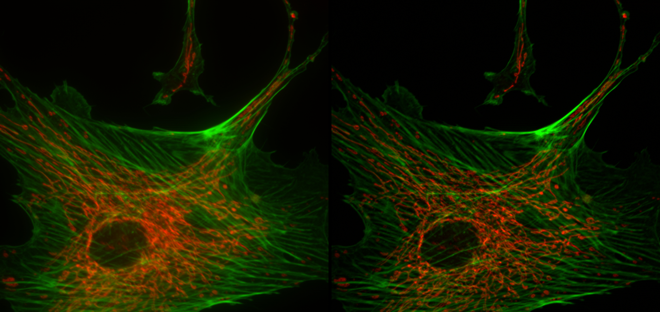
Standard Wide-Field Fluorescence Compared to Structured Illumination
Media Details
Created 3/7/2006 6:00:00 AM
In these images of bovine pulmonary artery endothelial cells, the mitochondria are stained with MitoTracker Red CMXRos (red), and F-actin is stained with BODIPY FL phallacidin (green). On the left is a standard wide-field fluorescence image that includes signal fluorescence from above and below focus. On the right is the same image following deconvolution using the Zeiss Apotome Structured Illumination System.
Credits
- Jon Ekman , ITG, Beckman Institute
Visualization Laboratory
Beckman Institute room 2203
405 North Mathews Avenue, Urbana, IL
(217) 300-0566