Image of the Week Gallery
Synapse Identification in a Mouse Visual Cortex
Media Details
Created 2/25/2003 6:00:00 AM
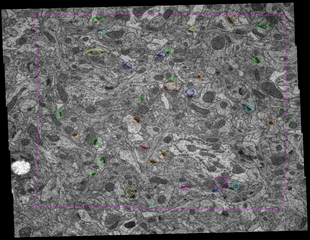
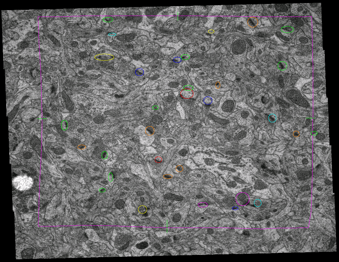
This image is of a transmission electron micrograph of the mouse visual cortex, acquired digitally using the ITG's Philips CM200, and tiled (5x5) using Teitz imaging software. This is the seventh of ten serial micrographs, and on each micrograph in the series, synapses were circled as they appeared (different color for each micrograph). This method allows for an unbiased estimate of synapse density in the neural tissue. Software for aligning micrographs and tracing synapses available at http://synapses.bu.edu/ and in the VMIL. This research is conducted under the advisement of Dr. William Greenough an Aaron W. Grossman.
Credits
- Hooma Khaliq , Beckman Institute
- Jessica Martellaro , Beckman Institute
- Brad Shufelt , Beckman Institute
- Aaron Grossman , Beckman Institute
- Nick Elisseou , Beckman Institute
- Cyrus Press , Beckman Institute
- Ken Schulz , Beckman Institute
- Ok-kyung Kim , Beckman Institute
- Loukas Koyonos , Beckman Institute
Visualization Laboratory
Beckman Institute room 2203
405 North Mathews Avenue, Urbana, IL
(217) 300-0566